Diagram Of An Animal Cell Under A Microscope - >animal cell microscope slide | wallpapersskin - Answer the following questions in your exercise book.
byNicky Didamo-
0
Diagram Of An Animal Cell Under A Microscope - >animal cell microscope slide | wallpapersskin - Answer the following questions in your exercise book.. Smooth endoplasmic reticulum, mitochondria, golgi bodies, lysosomes. (iii) presence of cell wall. Cells are covered by a cell membrane and come in many different shapes. (refer to box 7.1 on p. But at the same time it is interpretive.
Be careful pushing it under the clips that the cover slide doesn't move or crack. But at the same time it is interpretive. A scale bar has been marked on the drawing, allowing the. Click (or tap) the diagram for a simple labelled version. Most cells, both animal and plant, range in size between 1 and 100 micrometers and are thus visible only with the aid of a microscope.
Cells under a microscope : Biological Science Picture ... from pulpbits.net Here's a diagram of a plant cell: Line diagram of a general animal cell. Here's a photo of a plant cell under an electron microscope. Here's a photo of a plant cell under an electron microscope. Answer the following questions in your exercise book. If you examine plant and animal cells under a microscope you will note major structural differences between both. Removing cellular waste products from the cell. Most cells, both animal and plant, range in size between 1 and 100 micrometers and are thus visible only with the aid of a microscope.
Plant cells have cell walls, one large vacuole per cell, and chloroplasts, while animal cells will have a cell membrane only.
The cell is the basic unit of life. Made the first compound microscope and observed a slice of a cork oak tree. 4) cell membrane or plasma membrane. Cell membrane, nucleus, and cytoplasm with frog red blood cells. Animal cells are the basic unit of life in organisms of the kingdom animalia. To study the microscopic structures of human cheek cells under a compound microscope. After completing this section, you should know: Smooth endoplasmic reticulum, mitochondria, golgi bodies, lysosomes. But at the same time it is interpretive. The contents of a cell are. Which represents the same cell, seen under a light (optical) microscope at which processes are shown in the diagram and involve the cell surface membrane of the cell? In truth, there are still features of plant and animal cells we're only lately. The diagram is very clear, and labeled;
Cells under microscope foto sin derechos de autor. Given below is the diagram of a cell as seen under the microscope after having been placed in a solution A active transport and diffusion b diffusion and. After completing this section, you should know: Made the first compound microscope and observed a slice of a cork oak tree.
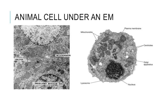
3. eukaryotes, their structure & em from image.slidesharecdn.com (iii) presence of cell wall. 4) cell membrane or plasma membrane. It is the outermost membrane of an animal cell having a thickness. Examining plant cells under the microscope. In fact, most are invisible without using a microscope. Made the first compound microscope and observed a slice of a cork oak tree. Under the microscope, an animal cell shows many different parts called organelles, that work together to keep the cell functional. (ii) presence of large central vacuole in plant cell.
To study the microscopic structures of human cheek cells under a compound microscope.
(iii) presence of cell wall. Support their own weight (which animals usually do by means of a skeleton). Cellular respiration occurs in mitochondria on animal cells, which are structurally somewhat analogous to chloroplasts, and also perform the function of producing energy. This draw and label the structure of a generalized animal cell (i.e. The diagram is very clear, and labeled the diagram is very clear, and labeled; Select the lowest power objective lens. Smooth endoplasmic reticulum, mitochondria, golgi bodies, lysosomes. A cell is a very tiny structure which exists in living bodies. • students will look at a blood smear to identify blood cells. Cell is a tiny structure and functional unit of a living organism containing various parts known as organelles. Students will observe cheek cells under a microscope. Be careful pushing it under the clips that the cover slide doesn't move or crack. Animal cells are the basic unit of life in organisms of the kingdom animalia.
Students will observe cheek cells under a microscope. You can specify conditions of storing and accessing cookies in. In truth, there are still features of plant and animal cells we're only lately. The diagram is very clear, and labeled; 17) microscopic examination of an animal cell reveals the presence of a plasma membrane but no cell wall which.
Biomimicry: A Soft 3D-Printed Seat Inspired by Plant Cell ... from i.pinimg.com Animal cells are the basic unit of life in organisms of the kingdom animalia. A active transport and diffusion b diffusion and. You can specify conditions of storing and accessing cookies in. A comparison of plant and animal cells using labelled diagrams and descriptive explanations. Be careful pushing it under the clips that the cover slide doesn't move or crack. All organisms are made up of cells (or in some cases, a single cell). Here's a diagram of a plant cell: Click (or tap) the diagram for a simple labelled version.
To study the microscopic structures of human cheek cells under a compound microscope.
Cells of plant or animal tissue. All organisms are made up of cells (or in some cases, a single cell). Plant cells have cell walls, one large vacuole per cell, and chloroplasts, while animal cells will have a cell membrane only. Learn about the similarities and differences between produce their own food (which they do in a process called photosynthesis). To study the microscopic structures of human cheek cells under a compound microscope. This is a diagram of a typical plant cell. The cell is the basic unit of life. Here's a diagram of a plant cell: The role and function of the plasma membrane; (iii) presence of cell wall. Answer the following questions in your exercise book. Under the microscope, an animal cell shows many different parts called organelles, that work together to keep the cell functional. To look at a cell close up we need a microscope.