Plant Cell Under Microscope 100X : Leaf Structure Under The Microscope / The best settings for viewing yeast on a microscope is a numerical aperture of at least 1.4, and a magnification of the objective lens at around 60x to 100x.
byNicky Didamo-
0
Plant Cell Under Microscope 100X : Leaf Structure Under The Microscope / The best settings for viewing yeast on a microscope is a numerical aperture of at least 1.4, and a magnification of the objective lens at around 60x to 100x.. Photo micro sections with high magnification with light microscope. The best settings for viewing yeast on a microscope is a numerical aperture of at least 1.4, and a magnification of the objective lens at around 60x to 100x. Besides good quality brands, you'll also find plenty of discounts when you shop for microscope 100x during big sales. The cut root of a young horse bean. Plant cell structure under microscope :
Some organelles are visible with a compound light microscope, while other organelles can be seen only plant cell organelles that are invisible under a compound light microscope include mitochondria, ribosomes, endoplasmic reticula, and golgi bodies. 3.8 out of 5 stars 175. It is a lateral meristematic. Chloroplast flowing inside the plant cell under 100x len. Use a scalpel to cut off a thin inner layer of an onion.
Plant Cells Under Microscope 100x Canstock from cdn.w600.comps.canstockphoto.com 1200 x 1600 jpeg 343 кб. Many educational facilities use the procedure as an experiment for students to explore the principles of microscopy and the identification of cells, and viewing cheek cells is one of. Data analysis cell shape solution. Micro photo of corn cells. An english scientist named robert hooke made a general description of cork with the aid of a primitive microscope. Find out more about plants under the microscope in this video from kew. Same as the plant cell, we can see general and important parts of the eukaryotic cell. Put the layer of onion above a microscope slide and apply dye on it.
Observing human cheek cells under a microscope is a simple way to quickly view and learn about human cell structure.
Plant cells under the microscope. Plant & animal cells staining lab answers | schoolworkhelper. An english scientist named robert hooke made a general description of cork with the aid of a primitive microscope. Transfer your mount to the microscope and examine the radicle under low power. Put the layer of onion above a microscope slide and apply dye on it. Dapi do not penetrate intact plant cells. Yeast can be viewed under the microscope through two different microscopy techniques. Chloroplast flowing inside the plant cell under 100x len. Plant cell under microscope labeledshow all. For the experiment you will only need onion, dropper and the microscope (container and tools are optional). How can you see a plant cell under a compound microscope? 1280 x 720 jpeg 75 кб. It is a lateral meristematic.
1280 x 720 jpeg 75 кб. In this experiment we will see onion cells under the microscope. Chloroplast flowing inside the plant cell under 100x len. Green plant cells under microscope seamless vector pattern. Same as the plant cell, we can see general and important parts of the eukaryotic cell.
Plant Cells Under Microscope 100x Canstock from comps.canstockphoto.com A cell is a very tiny structure which exists in living bodies. Plant cell under microscope with 100x magnification power. Under the electron microscope here you can see. Some organelles are visible with a compound light microscope, while other organelles can be seen only plant cell organelles that are invisible under a compound light microscope include mitochondria, ribosomes, endoplasmic reticula, and golgi bodies. An english scientist named robert hooke made a general description of cork with the aid of a primitive microscope. Plant cells under the microscope. Plant cell structure under microscope : Plant cells from flower petal under microscope.
Find out more about plants under the microscope in this video from kew.
Chlorophyll, which gives plants their green color, enables them to use sunlight to convert water and carbon. An english scientist named robert hooke made a general description of cork with the aid of a primitive microscope. Cork or cork cambium (pl. Boletus fruiting body under the microscope 100x across. Preparation of onion cell slide and viewing under a light microscope. Green plant cells under microscope seamless vector pattern. Micro photo of corn cells. The systems work together to produce a magnified image of the specimen under examination. Transfer your mount to the microscope and examine the radicle under low power. Plant cell under microscope labeledshow all. A cell is a very tiny structure which exists in living bodies. Chloroplast flowing inside the plant cell under 100x len of a microscope. Observing human cheek cells under a microscope is a simple way to quickly view and learn about human cell structure.
In this experiment we will see onion cells under the microscope. Same as the plant cell, we can see general and important parts of the eukaryotic cell. This value corresponds to the number of electrons received by the detector during a small period of time of the scanning when the beam is targeted to the (x, y) electron microscopy of plant cells. How can you see a plant cell under a compound microscope? Micro photo of corn cells.
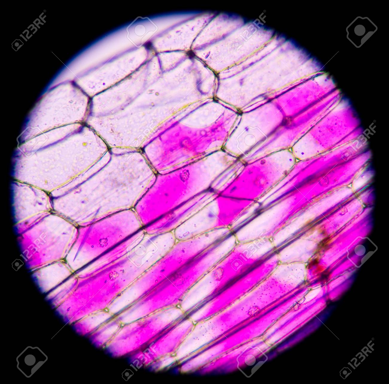
Pink Plant Cells Under Microscope 400x Stock Photo Picture And Royalty Free Image Image 43886014 from previews.123rf.com The best settings for viewing yeast on a microscope is a numerical aperture of at least 1.4, and a magnification of the objective lens at around 60x to 100x. The systems work together to produce a magnified image of the specimen under examination. Looking for a good deal on microscope 100x? Data analysis cell shape solution. The beginner's guide to microscopy. It is a lateral meristematic. Some organelles are visible with a compound light microscope, while other organelles can be seen only plant cell organelles that are invisible under a compound light microscope include mitochondria, ribosomes, endoplasmic reticula, and golgi bodies. Plant cell structure under microscope :
In this experiment we will see onion cells under the microscope.
You can see the veins of it under 100x magnification! Looking for a good deal on microscope 100x? Shed mule from waterloo structures. The majority of sections that you will be given to look at in the virtual plant exercises, will have been cut in the transverse plane. Same as the plant cell, we can see general and important parts of the eukaryotic cell. This value corresponds to the number of electrons received by the detector during a small period of time of the scanning when the beam is targeted to the (x, y) electron microscopy of plant cells. The best settings for viewing yeast on a microscope is a numerical aperture of at least 1.4, and a magnification of the objective lens at around 60x to 100x. Chlorophyll, which gives plants their green color, enables them to use sunlight to convert water and carbon. 1200 x 1600 jpeg 343 кб. The cut root of a young horse bean. Some organelles are visible with a compound light microscope, while other organelles can be seen only plant cell organelles that are invisible under a compound light microscope include mitochondria, ribosomes, endoplasmic reticula, and golgi bodies. For the experiment you will only need onion, dropper and the microscope (container and tools are optional). Chloroplast flowing inside the plant cell under 100x len.